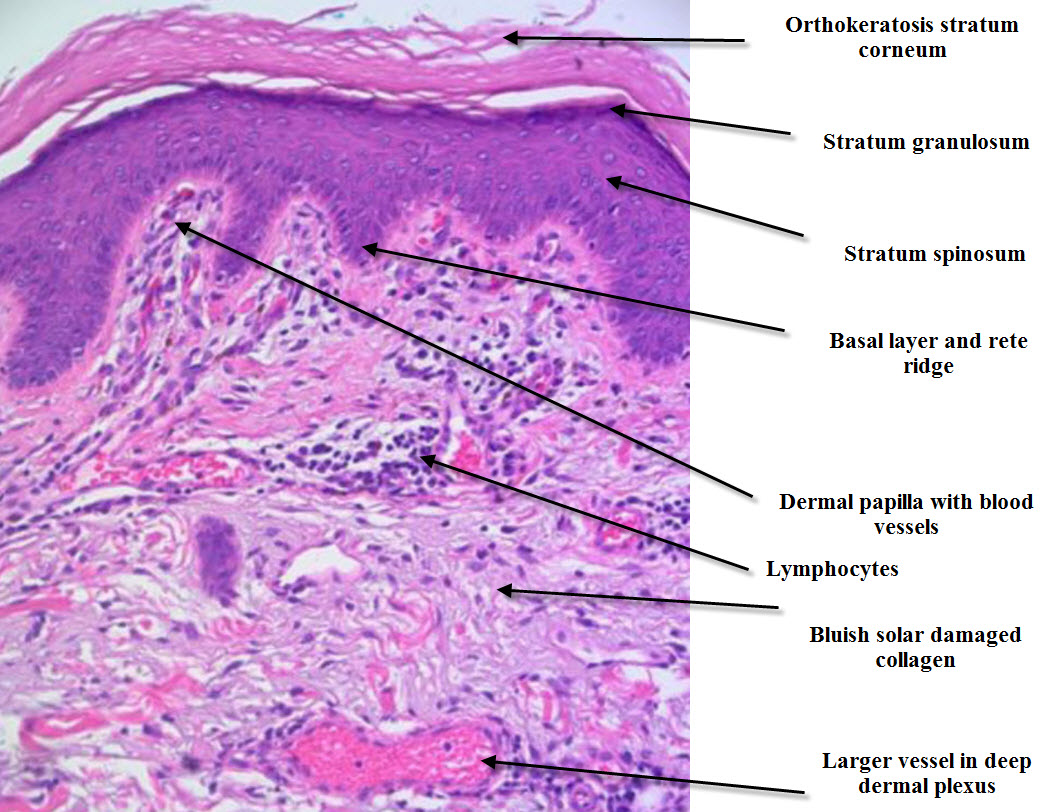
Histologic Layers Of Skin
Histology drawings: january 2014 Skin stratum palm function structure corneum eosinophilic separating clearly overlying granular lucidum layer fig cell note Chemical burns
Histology Drawings: January 2014
Histology dermis epithelial sebaceous physiology glands membrane corpuscles appendages krause zapisano receptors Histologic "onion skin" appearance characterized by concentric layers Histologic layers concentric characterized
Histology of skin
Histology (skin)The structure and function of skin Skin (integumentary system)Skin histology.
Histology nus pathweb annotations expandSkin histopathology introduction simple dermatopathology made dermpath inflammatory Histology skin thin system integumentary human anatomy thick drawings section cross mallory slides trichrome nervous cutis renal 40x between bodyHistology skin.

Histologic tissue eosin staining hematoxylin
Skin – normal histology – nus pathweb :: nus pathwebHistology of the skin Skin osmosis histology continue learningHistologic evaluation of skin tissue by hematoxylin and eosin staining.
Skin layers anatomy chemical epidermis structure levels peel layer dermis microscopic section peeling peels epidermal bestofbothworldsaz stratum cross tissue corneumHistology integumentary trichrome mallory physiology facmedicine eosin hematoxylin cutis Dermatopathology made simpleSkin thin histology thick integumentary system drawings slides human hematoxylin eosin trichrome through biology reference links.

Layers histologic skin burns chemical permission reproduced
.
.


